
Page 30 - The Beauty and Sorrow in Endodontics-Chapter 1
P. 30
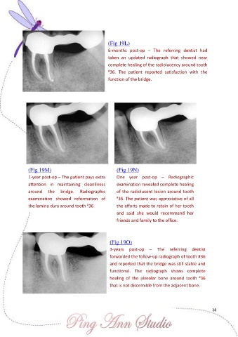
(Fig 19L)

                                                       6-months  post-op  –  The  referring  dentist  had
                                                       taken  an  updated  radiograph  that  showed  near

                                                       complete healing of the radiolucency around tooth
                                                       # 36.  The  patient  reported  satisfaction  with  the
                                                       function of the bridge.


               (Fig 19M)                                   (Fig 19N)
               1-year post-op – The patient pays extra     One  year  post-op  –  Radiographic

               attention  in  maintaining  cleanliness     examination revealed complete healing
               around  the     bridge.  Radiographic       of the radiolucent lesion around tooth
               examination  showed  reformation  of        # 36. The patient was appreciative of all
               the lamina dura around tooth  36.           the efforts made to retain of her tooth
                                             #
                                                           and  said  she  would  recommend  her
                                                           friends and family to the office.


                                                        (Fig 19O)
                                                        3-years  post-op  –  The  referring  dentist

                                                        forwarded the follow-up radiograph of tooth #36
                                                        and reported that the bridge was still stable and
                                                        functional.  The  radiograph  shows  complete
                                                                                                     #
                                                        healing  of  the  alveolar  bone  around  tooth  36
                                                        that is not discernible from the adjacent bone.


                                                                                                           28