
Page 29 - The Beauty and Sorrow in Endodontics-Chapter 1
P. 29
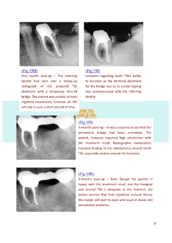
(Fig 19H)                                   (Fig 19I)
                One  month  post-op  –  The  referring      Concerns  regarding  tooth  38’s  ability
                                                                                       #
                dentist  had  sent  over  a  follow-up      to  function  as  the  terminal  abutment
                radiograph  of  the  prepared      # 36     for the bridge due to its mesial tipping

                abutment  with  a  temporary  36-x-38       was  communicated  with  the  referring
                bridge. The patient was ecstatic to have    dentist.
                regained  masticatory  function  on  the
                left side in such a short period of time.


                                                       (Fig 19J)
                                                       3-months post-op – It was a surprise to see that the

                                                       permanent  bridge  had  been  cemented.  The
                                                       patient,  however,  reported  high  satisfaction  with
                                                       the  treatment  result.  Radiographic  examination

                                                       revealed healing of the radiolucency around tooth
                                                       # 36, especially evident around the furcation.


                                                       (Fig 19K)

                                                       3-months  post-op  –  Even  though  the  patient  is
                                                       happy  with  the  treatment  result  and  the  marginal
                                                                    #
                                                       seal  around  38  is  adequate  at  the  moment,  the
                                                       author worries that from repetitive occlusal forces,
                                                       the margin will start to open and result in decay and
                                                       periodontal problems.


                                                                                                           27