
Page 28 - The Beauty and Sorrow in Endodontics-Chapter 1
P. 28
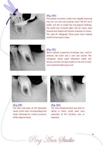
(Fig 19D)

                                                       The patient returned a week later happily reporting
                                                                                              #
                                                       that  she  can  now  eat  properly,  tooth  36  felt  more
                                                       stable, and she no longer has any gingival swellings.

                                                       The  tooth  was  accessed  again  and  all  canals  were
                                                       cleaned and shaped utilizing the envelope of motion.
                                                       The  cone  fit  radiograph  shows  good  canal  shaping
                                                       and fit of the master cones.


                                                       (Fig 19E)
                                                       Warm  vertical  compaction  technique  was  used  to

                                                       obturate  the  tooth  and  a  core  was  placed.  The
                                                       radiograph  shows  good  obturation  depth  and
                                                       density, and the extruded sealers at the exit of each
                                                       canal represent tight apical seal.


                (Fig 19F)                                   (Fig 19G)
                The  flow  and  taper  of  the  obturated   The  only  disappointment  was  that  no
                canals match their corresponding root       lateral  or  furcal  canals  were  seen
                shape  following  the  natural  curvature   obturated  at  the  furcation  area  as

                of the original canals.                     expected.


                                                                                                           26