
Page 25 - The Beauty and Sorrow in Endodontics-Chapter 1
P. 25
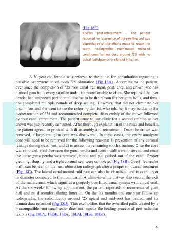
(Fig 18F)

                                                          4-years  post-retreatment  –  The  patient
                                                          reported no recurrence of the swelling and was
                                                          appreciative of the efforts made to retain the
                                                          tooth.  Radiographic  examination  revealed
                                                                                           #
                                                          continuous  lamina  dura  around  25  with  no
                                                          apical radiolucency or signs of infection.


                     A 30-year-old female was referred to the clinic for consultation regarding a
                                                 #
            possible overextension  of  tooth  25  obturation  (Fig  18A).  According  to  the patient,
                                             #
            ever since the completion of  25 root canal treatment, post, core, and crown, she has
            noticed gum boils every so often and it is uncomfortable to chew. She reported that her

            dentist had suspected periodontal disease to be the reason for her gum boils, and thus,
            has completed multiple rounds of deep scaling. However, that did not eliminate her
            discomfort and she went to see the referring dentist, who told her it may be due to the
                                #
            overextension of  25 and recommended complete disassembly of the crown followed
            by root canal retreatment. The patient came to our clinic for a second opinion as her
            crown was just recently cemented. After thorough explanation of the risks and benefits,
            the patient agreed to proceed with disassembly and retreatment. Once the crown was
            removed, a large amalgam core was discovered. In these cases, the entire amalgam

            core will need to be removed for the following reasons: 1) prevention of any coronal
            leakage during treatment, and 2) to assess the remaining tooth structure. Once the core
            was removed, voids between the gutta percha and dentin wall were observed, and once

            the loose gutta percha was removed, blood and pus gushed out of the canal.  Proper
            cleaning, shaping, and a tight coronal seal were completed (Fig 18B). Overfilled sealer
            puffs can be seen on the post-obturation radiograph after a proper root canal treatment
            (Fig 18C). The lateral canal around mid-root can also be visualized and is even larger
            in diameter compared to the main canal. A white-in-white dotwas also seen at the exit

            of the main canal, which signifies a properly overfilled canal system with apical seal.
            At the six-weeks follow-up appointment, the patient reported no recurrence of gum
            boil  and  no  discomfort  during  function.  On  the  six-months  and  one-year  follow-up
                                                         #
            radiographs,  the  radiolucency  around  25  apical  and  mid-root  has  healed,  and  its
            lamina dura reformed (Fig 18D). This exemplifies that the overfilled puffs created by a
            biocompatible root canal sealer does not impede the healing process of peri-radicular
            lesions (Fig 18E/a, 18E/b, 18E/c, 18E/d, 18E/e, 18E/f).


                                                                                                           23