
Page 22 - The Beauty and Sorrow in Endodontics-Chapter 1
P. 22
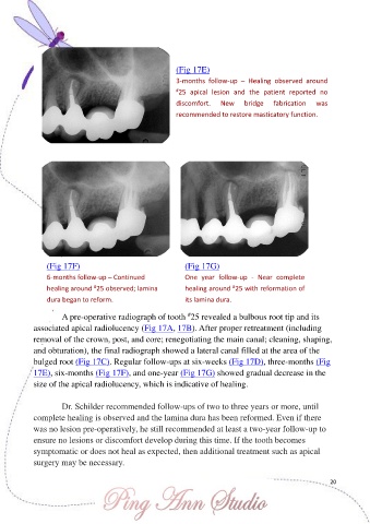
(Fig 17E)

                                                          3-months  follow-up  –  Healing  observed  around
                                                          # 25  apical  lesion  and  the  patient  reported  no
                                                          discomfort.   New     bridge   fabrication   was
                                                          recommended to restore masticatory function.


                (Fig 17F)                                    (Fig 17G)

                6-months follow-up – Continued               One  year  follow-up  -  Near  complete
                               #
                                                                            #
                healing around  25 observed; lamina          healing around  25 with reformation of
                dura began to reform.                        its lamina dura.


                                                             #
                     A pre-operative radiograph of tooth  25 revealed a bulbous root tip and its
            associated apical radiolucency (Fig 17A, 17B). After proper retreatment (including
            removal of the crown, post, and core; renegotiating the main canal; cleaning, shaping,
            and obturation), the final radiograph showed a lateral canal filled at the area of the

            bulged root (Fig 17C). Regular follow-ups at six-weeks (Fig 17D), three-months (Fig
            17E), six-months (Fig 17F), and one-year (Fig 17G) showed gradual decrease in the
            size of the apical radiolucency, which is indicative of healing.


                     Dr. Schilder recommended follow-ups of two to three years or more, until
            complete healing is observed and the lamina dura has been reformed. Even if there
            was no lesion pre-operatively, he still recommended at least a two-year follow-up to
            ensure no lesions or discomfort develop during this time. If the tooth becomes

            symptomatic or does not heal as expected, then additional treatment such as apical
            surgery may be necessary.


                                                                                                           20