
Page 20 - The Beauty and Sorrow in Endodontics-Chapter 1
P. 20
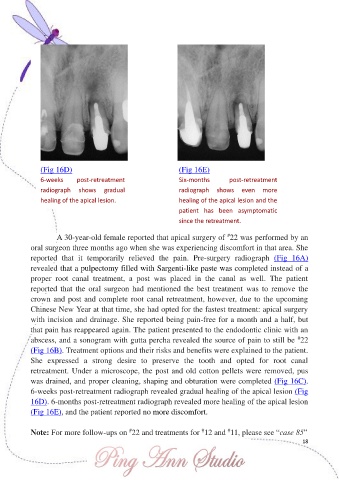
(Fig 16D)                                         (Fig 16E)

               6-weeks      post-retreatment                    Six-months        post-retreatment

               radiograph  shows  gradual                       radiograph  shows  even  more
               healing of the apical lesion.                    healing of the apical lesion and the

                                                                patient  has  been  asymptomatic
                                                                since the retreatment.

                                                                                 #
                     A 30-year-old female reported that apical surgery of  22 was performed by an
            oral surgeon three months ago when she was experiencing discomfort in that area. She

            reported  that  it  temporarily  relieved  the  pain.  Pre-surgery  radiograph  (Fig  16A)
            revealed that a pulpectomy filled with Sargenti-like paste was completed instead of a
            proper  root  canal  treatment,  a  post  was  placed  in  the  canal  as  well.  The  patient
            reported that  the  oral surgeon had  mentioned the best  treatment  was  to remove the
            crown and post and complete root canal retreatment, however, due to the upcoming

            Chinese New Year at that time, she had opted for the fastest treatment: apical surgery
            with incision and drainage. She reported being pain-free for a month and a half, but
            that pain has reappeared again. The patient presented to the endodontic clinic with an

                                                                                                          #
            abscess, and a sonogram with gutta percha revealed the source of pain to still be  22
            (Fig 16B). Treatment options and their risks and benefits were explained to the patient.
            She  expressed  a  strong  desire  to  preserve  the  tooth  and  opted  for  root  canal
            retreatment. Under a microscope, the post and old cotton pellets were removed, pus
            was drained, and proper cleaning, shaping and obturation were completed (Fig 16C).

            6-weeks post-retreatment radiograph revealed gradual healing of the apical lesion (Fig
            16D). 6-months post-retreatment radiograph revealed more healing of the apical lesion
            (Fig 16E), and the patient reported no more discomfort.


                                                                                 #
                                                                        #
                                              #
            Note: For more follow-ups on  22 and treatments for  12 and  11, please see “case 85”
                                                                                                           18