
Page 15 - The Beauty and Sorrow in Endodontics-Chapter 1
P. 15
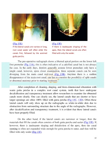
(Fig 13E)                                    (Fig 13F)
               If the lateral canals are narrow or long,    If  there  is  inadequate  shaping  of  the

               root  canal  sealer  will  often  enter  the   apex, then the lateral canals are often
               canals  first,  followed  by  the  warmed    filled with only the sealer.

               gutta percha.

                     The pre-operative radiograph shows a blurred apical portion on the lower left
            first premolar (Fig 13A), this is often indicative of a calcified canal but is not always
            the  case.  In  the  early  days,  dentists  generally  assume  lower  premolars  only  have  a

            single  canal,  however,  upon  closer  examination,  three  separate  canals  can  be  seen
            diverging  from  the  main  canal  mid-root  (Fig  13B).  Anytime  there  is  a  sudden
            disappearance of the main root canal, one has to consider the possibility of split canals

            or abnormal anatomy prior to starting treatment.

                     After completion of cleaning, shaping, and three-dimensional obturation with
            warm  gutta  percha  in  a  complex  root  canal  system,  teeth  that  have  undergone
            decalcification and transparency treatment allow researchers to examine the obturated

            canals  more clearly. One can  clearly  see, the  lateral canals  that are  shorter  or have
            larger openings are often 100% filled with gutta percha (Fig 13C, 13D). Often these
            lateral  canals  will  only  show  up  on  the  radiographs  as  white-in-white  dots  due  to

            obstruction from surrounding structure due to the angle of the radiographs. However,
            after decalcification and transparency treatment, it is evident that these lateral canals
            have been properly filled.

                     On  the  other  hand,  if  the  lateral  canals  are  narrower  or  longer,  then  the

            materials that fill the canals often consists of both gutta percha and sealer (Fig 13E). If,
            however,  there  is  inadequate  shaping  and  disinfection  of  the  apex,  then  the  canal
            opening is often not expanded wide enough for gutta percha to enter, and thus will be

            filled with only sealer (Fig 13F).

                                                                                                           13