
Page 11 - The Beauty and Sorrow in Endodontics-Chapter 1
P. 11
Case 11
Completion of proper root canal treatment will sometimes
reveal lateral canals that could not have been predicted
prior to treatment
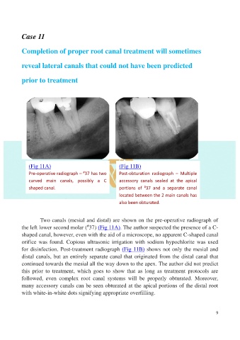
(Fig 11A) (Fig 11B)
#
Pre-operative radiograph – 37 has two Post-obturation radiograph – Multiple
curved main canals, possibly a C accessory canals sealed at the apical
#
shaped canal. portions of 37 and a separate canal
located between the 2 main canals has
also been obturated.
Two canals (mesial and distal) are shown on the pre-operative radiograph of
the left lower second molar ( 37) (Fig 11A). The author suspected the presence of a C-
#
shaped canal, however, even with the aid of a microscope, no apparent C-shaped canal
orifice was found. Copious ultrasonic irrigation with sodium hypochlorite was used
for disinfection. Post-treatment radiograph (Fig 11B) shows not only the mesial and
distal canals, but an entirely separate canal that originated from the distal canal that
continued towards the mesial all the way down to the apex. The author did not predict
this prior to treatment, which goes to show that as long as treatment protocols are
followed, even complex root canal systems will be properly obturated. Moreover,
many accessory canals can be seen obturated at the apical portions of the distal root
with white-in-white dots signifying appropriate overfilling.
9