
Page 13 - The Beauty and Sorrow in Endodontics-Chapter 1
P. 13
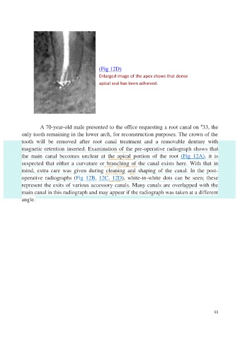
(Fig 12D)

                                                  Enlarged image of the apex shows that dense
                                                  apical seal has been achieved.


                                                                                                     #
                     A 70-year-old male presented to the office requesting a root canal on  33, the
            only tooth remaining in the lower arch, for reconstruction purposes. The crown of the
            tooth  will  be  removed  after  root  canal  treatment  and  a  removable  denture  with
            magnetic retention inserted. Examination of the pre-operative radiograph shows that

            the  main  canal  becomes  unclear  at  the  apical  portion  of  the  root  (Fig  12A),  it  is
            suspected that either a curvature or branching of the canal exists here. With that in
            mind,  extra  care  was  given  during  cleaning  and  shaping  of  the  canal.  In  the  post-
            operative  radiographs  (Fig  12B,  12C,  12D),  white-in-white  dots  can  be  seen;  these
            represent the exits of various accessory canals. Many canals are overlapped with the

            main canal in this radiograph and may appear if the radiograph was taken at a different
            angle.


                                                                                                           11