
Page 35 - The Beauty and Sorrow in Endodontics-Chapter 1
P. 35
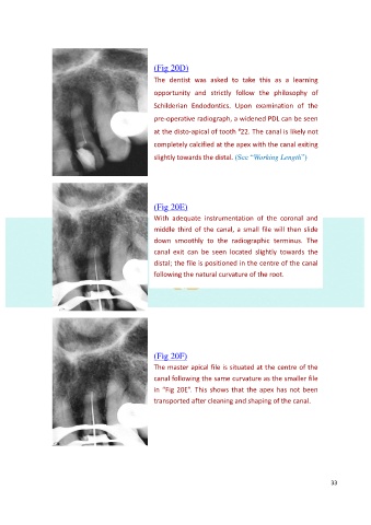
(Fig 20D)
                                                  The  dentist  was  asked  to  take  this  as  a  learning

                                                  opportunity  and  strictly  follow  the  philosophy  of
                                                  Schilderian  Endodontics.  Upon  examination  of  the

                                                  pre-operative radiograph, a widened PDL can be seen

                                                                             #
                                                  at the disto-apical of tooth  22. The canal is likely not
                                                  completely calcified at the apex with the canal exiting

                                                  slightly towards the distal. (See “Working Length”)


                                                  (Fig 20E)

                                                  With  adequate  instrumentation  of  the  coronal  and
                                                  middle third of the canal, a small file will then slide

                                                  down  smoothly  to  the  radiographic  terminus.  The
                                                  canal  exit  can  be  seen  located  slightly  towards  the
                                                  distal; the file is positioned in the centre of the canal
                                                  following the natural curvature of the root.


                                                  (Fig 20F)

                                                  The master apical file is situated at the centre of the
                                                  canal following the same curvature as the smaller file

                                                  in “Fig 20E”. This shows that the apex has not been
                                                  transported after cleaning and shaping of the canal.


                                                                                                           33