
Page 37 - The Beauty and Sorrow in Endodontics-Chapter 1
P. 37
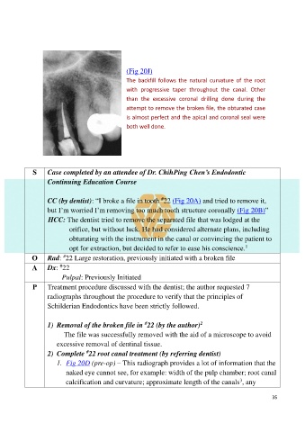
(Fig 20J)

                                                  The  backfill  follows  the  natural  curvature  of  the  root
                                                  with  progressive  taper  throughout  the  canal.  Other
                                                  than  the  excessive  coronal  drilling  done  during  the

                                                  attempt to remove the broken file, the obturated case
                                                  is almost perfect and the apical and coronal seal were
                                                  both well done.


              S     Case completed by an attendee of Dr. ChihPing Chen’s Endodontic
                    Continuing Education Course


                    CC (by dentist): “I broke a file in tooth  22 (Fig 20A) and tried to remove it,
                                                                #
                    but I’m worried I’m removing too much tooth structure coronally (Fig 20B)”
                    HCC: The dentist tried to remove the separated file that was lodged at the

                            orifice, but without luck. He had considered alternate plans, including
                            obturating with the instrument in the canal or convincing the patient to
                                                                                                 1
                            opt for extraction, but decided to refer to ease his conscience.
                          #
              O     Rad:  22 Large restoration, previously initiated with a broken file
                        #
              A     Dx:  22
                         Pulpal: Previously Initiated

              P     Treatment procedure discussed with the dentist; the author requested 7
                    radiographs throughout the procedure to verify that the principles of

                    Schilderian Endodontics have been strictly followed.

                                                                               2
                                                          #
                    1)  Removal of the broken file in  22 (by the author)
                           The file was successfully removed with the aid of a microscope to avoid
                       excessive removal of dentinal tissue.
                                   #
                    2)  Complete  22 root canal treatment (by referring dentist)
                       1.  Fig 20D (pre-op) – This radiograph provides a lot of information that the

                           naked eye cannot see, for example: width of the pulp chamber; root canal
                                                                                               3
                           calcification and curvature; approximate length of the canals , any

                                                                                                           35