
Page 22 - The Beauty and Sorrow in Endodontics-Prologue
P. 22
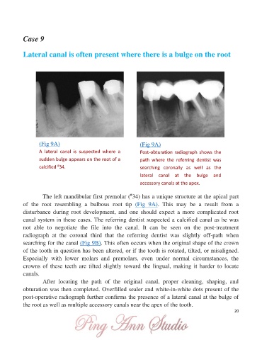
Case 9


            Lateral canal is often present where there is a bulge on the root


                   (Fig 9A)                                       (Fig 9A)

                   A  lateral  canal  is  suspected  where  a     Post-obturation  radiograph  shows  the
                   sudden bulge appears on the root of a          path  where  the  referring  dentist  was
                   calcified  34.                                 searching  coronally  as  well  as  the
                            #

                                                                  lateral  canal  at  the  bulge  and

                                                                  accessory canals at the apex.

                                                              #
                     The left mandibular first premolar ( 34) has a unique structure at the apical part
            of  the  root  resembling  a  bulbous  root  tip  (Fig  9A).  This  may  be  a  result  from  a

            disturbance  during  root  development,  and  one  should  expect  a  more  complicated  root
            canal system in these cases. The referring dentist suspected a calcified canal as he was
            not  able  to  negotiate  the  file  into  the  canal.  It  can  be  seen  on  the  post-treatment

            radiograph  at  the  coronal  third  that  the  referring  dentist  was  slightly  off-path  when
            searching for the canal (Fig 9B). This often occurs when the original shape of the crown
            of the tooth in question has been altered, or if the tooth is rotated, tilted, or misaligned.
            Especially  with  lower  molars  and  premolars,  even  under  normal  circumstances,  the
            crowns  of  these  teeth  are  tilted  slightly  toward  the  lingual,  making  it  harder  to  locate

            canals.
                     After  locating  the  path  of  the  original  canal,  proper  cleaning,  shaping,  and
            obturation was then completed. Overfilled sealer and white-in-white dots present of the

            post-operative radiograph further confirms the presence of a lateral canal at the bulge of
            the root as well as multiple accessory canals near the apex of the tooth.
                                                                                                               20