
Page 14 - The Beauty and Sorrow in Endodontics-Prologue
P. 14
Case 4
Radiographs taken at different angles often provide new insight
to the case
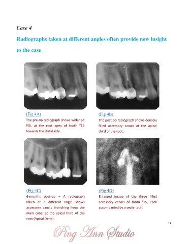
(Fig 4A) (Fig 4B)
The pre-op radiograph shows widened The post-op radiograph shows densely
#
PDL at the root apex of tooth 15 filled accessory canals at the apical
towards the distal side. third of the root.
(Fig 4C) (Fig 4D)
6-months post-op – A radiograph Enlarged image of the three filled
#
taken at a different angle shows accessory canals of tooth 15, each
accessory canals branching from the accompanied by a sealer puff.
main canal at the apical third of the
root (Apical Delta).
12