
Page 13 - The Beauty and Sorrow in Endodontics-Prologue
P. 13
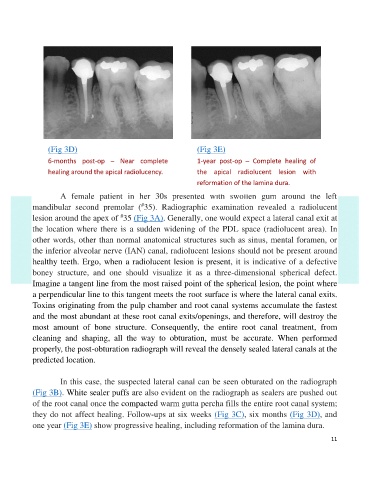
(Fig 3D)                                         (Fig 3E)
                 6-months  post-op  –  Near  complete             1-year  post-op  –  Complete  healing  of

                 healing around the apical radiolucency.          the  apical  radiolucent  lesion  with
                                                                  reformation of the lamina dura.

                     A  female  patient  in  her  30s  presented  with  swollen  gum  around  the  left
                                                #
            mandibular  second  premolar  ( 35).  Radiographic  examination  revealed  a  radiolucent
                                         #
            lesion around the apex of  35 (Fig 3A). Generally, one would expect a lateral canal exit at
            the location where there is a sudden widening of the PDL space (radiolucent area).  In
            other words, other than normal anatomical structures such as sinus, mental foramen, or

            the inferior alveolar nerve (IAN) canal, radiolucent lesions should not be present around
            healthy teeth. Ergo, when a radiolucent lesion is present, it is indicative of a defective
            boney  structure,  and  one  should  visualize  it  as  a  three-dimensional  spherical  defect.

            Imagine a tangent line from the most raised point of the spherical lesion, the point where
            a perpendicular line to this tangent meets the root surface is where the lateral canal exits.
            Toxins originating from the pulp chamber and root canal systems accumulate the fastest
            and the most abundant at these root canal exits/openings, and therefore, will destroy the
            most  amount  of  bone  structure.  Consequently,  the  entire  root  canal  treatment,  from

            cleaning  and  shaping,  all  the  way  to  obturation,  must  be  accurate.  When  performed
            properly, the post-obturation radiograph will reveal the densely sealed lateral canals at the
            predicted location.


                     In this case, the suspected lateral canal can be seen obturated on the radiograph
            (Fig 3B). White sealer puffs are also evident on the radiograph as sealers are pushed out
            of the root canal once the compacted warm gutta percha fills the entire root canal system;
            they do not affect healing. Follow-ups at six weeks (Fig 3C), six months (Fig 3D), and

            one year (Fig 3E) show progressive healing, including reformation of the lamina dura.

                                                                                                               11