
Page 11 - The Beauty and Sorrow in Endodontics (Chapter 4 - Part 2)
P. 11
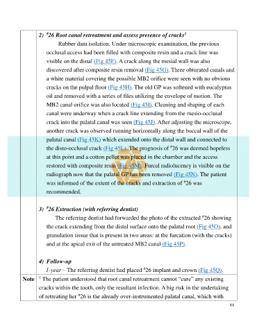
2)  26 Root canal retreatment and assess presence of cracks
                       #
                                                                                         1
                             Rubber dam isolation. Under microscopic examination, the previous

                       occlusal access had been filled with composite resin and a crack line was
                       visible on the distal (Fig 45F). A crack along the mesial wall was also

                       discovered after composite resin removal (Fig 45G). Three obturated canals and
                       a white material covering the possible MB2 orifice were seen with no obvious

                       cracks on the pulpal floor (Fig 45H). The old GP was softened with eucalyptus

                       oil and removed with a series of files utilizing the envelope of motion. The
                       MB2 canal orifice was also located (Fig 45I). Cleaning and shaping of each

                       canal were underway when a crack line extending from the mesio-occlusal
                       crack into the palatal canal was seen (Fig 45J). After adjusting the microscope,

                       another crack was observed running horizontally along the buccal wall of the
                       palatal canal (Fig 45K) which extended onto the distal wall and connected to

                       the disto-occlusal crack (Fig 45L). The prognosis of  26 was deemed hopeless
                                                                                  #
                       at this point and a cotton pellet was placed in the chamber and the access
                       restored with composite resin (Fig 45M). Furcal radiolucency is visible on the

                       radiograph now that the palatal GP has been removed (Fig 45N). The patient
                                                                                         #
                       was informed of the extent of the cracks and extraction of  26 was
                       recommended.


                       #
                    3)  26 Extraction (with referring dentist)
                             The referring dentist had forwarded the photo of the extracted  26 showing
                                                                                                 #
                       the crack extending from the distal surface onto the palatal root (Fig 45O), and

                       granulation tissue that is present in two areas: at the furcation (with the cracks)

                       and at the apical exit of the untreated MB2 canal (Fig 45P).


                    4)  Follow-up
                                                                     #
                       1-year – The referring dentist had placed  26 implant and crown (Fig 45Q).
                    1
            Note   The patient understood that root canal retreatment cannot “cure” any existing
                    cracks within the tooth, only the resultant infection. A big risk in the undertaking

                                      #
                    of retreating her  26 is the already over-instrumented palatal canal, which with
                                                                                                              44