
Page 15 - The Beauty and Sorrow in Endodontics-Chapter 3
P. 15
+
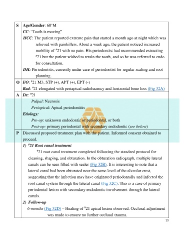
S Age/Gender: 60 M
CC: “Tooth is moving”
HCC: The patient reported extreme pain that started a month ago at night which was
relieved with painkillers. About a week ago, the patient noticed increased
mobility of 21 with no pain. His periodontist had recommended extracting
#
# 21 but the patient wished to retain the tooth, and so he was referred to endo
for consultation.
DH: Periodontitis, currently under care of periodontist for regular scaling and root
planning.
O I/O: 21 M3, STP (+), APT (+), EPT (-)
#
#
Rad: 21 elongated with periapical radiolucency and horizontal bone loss (Fig 32A)
A Dx: 21
#
Pulpal: Necrosis
Periapical: Apical periodontitis
Etiology:
Pre-op: unknown endodontic or periodontal, or both
Post-op: primary periodontal with secondary endodontic (see below)
P Discussed proposed treatment plan with the patient. Informed consent obtained to
proceed.
#
1) 21 Root canal treatment
# 21 root canal treatment completed following the standard protocol for
cleaning, shaping, and obturation. In the obturation radiograph, multiple lateral
canals can be seen filled with sealer (Fig 32B). It is interesting to note that a
lateral canal had been obturated near the same level of the alveolar crest,
suggesting that the infection may have originated periodontally and infected the
root canal system through the lateral canal (Fig 32C). This is a case of primary
periodontal lesion with secondary endodontic involvement through the lateral
canals.
2) Follow-up
#
6-months (Fig 32D) – Healing of 21 apical lesion observed. Occlusal adjustment
was made to ensure no further occlusal trauma.
13