
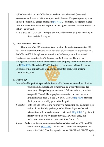
Page 15 - The Beauty and Sorrow in Endodontics-Chapter 4 - Part 1
P. 15
with ultrasonics and NaOCl solution to clean the split canal. Obturated

                    completed with warm vertical compaction technique. The post-op radiograph

                    showed both apical canals obturated (Fig 41D). Temporary restoration placed
                    and rubber dam removed. Post-op instructions given and the patient informed to

                    return in one week.
                   3-days post-op – Care call – The patient reported no more gingival swelling or

                                fever and she feels great.


                    #
                2)  34 Root canal treatment
                                                                                                     #
                                           #
                         One week after  35 retreatment completion, the patient returned for  34
                    root canal treatment. Intraoral exam revealed slight tenderness to percussion at
                         #
                    both  34 and  35, though not as sensitive as before anymore. Root canal
                                   #
                                                    #
                    treatment was completed on  34 under standard protocol. The post-op
                    radiograph showeda curved main canal with a properly filled lateral canal as

                    well (Fig 41E). The original  34- 35 splinted crowns were adjusted to prevent
                                                    #
                                                         #
                    excess occlusal contacts and temporarily cemented back. Oral hygiene
                    instructions given.


                3)  Follow-up

                   3-months –The patient reported she is now able to resume normal masticatory
                                function on both teeth and experienced no discomfort since the

                                                                            #
                                treatments. The probing depths around  35 has reduced to 3-5mm
                                              +
                                (originally 7 mm). Radiographic examination showed signs of
                                                                 #
                                                        #
                                healing around both  34 and  35 apical lesions (Fig 41F). Stressed
                                the important of oral hygiene with the patient.
                                                #
                                       #
                   6-months – Both  34 and  35 respond normally to percussion and palpation tests
                                and exhibited healthy probing depths. The radiograph showed
                                reformation of lamina dura around both teeth (Fig 41G). Significant

                                improvement in oral hygiene observed. New post, core, and
                                                                                         #
                                                                                #
                                individual crowns were recommended for  34 and  35.
                                                                                              #
                                                                                                       #
                   1-year – Radiographic examination revealed completed healing of  34 and  35
                                apical lesions (Fig 41H). The restoring dentist had completed the
                                                                                             #
                                                                                                  #
                                             #
                                                                                #
                                                  #
                                                                                    #
                                crowns for  34- 37 but has opted to splint  34- 35 and  36- 37 again.
                                                                                                           13